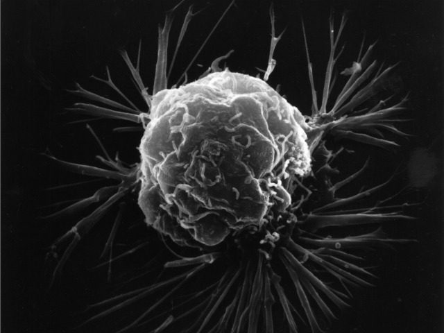
Researchers at UCLA have invented a method using a microscope augmented by artifical intelligence to detect cancer cells quicker and more accurately than current techniques.
Using a combination of photonic time stretch and deep learning, the device is capable of analyzing 36 million images per second while leaving blood samples undamaged so they may be used for other testing purposes.
The new photonic time stretch technology, invented by Barham Jalali who also led the study, takes pictures of blood cells using flashing lasers occurring in nanosecond bursts.
“Each frame is slowed down in time and optically amplified so it can be digitized,” said Ata Mahjoubfar, a UCLA postdoctoral fellow who worked on the project. “This lets us perform fast cell imaging that the artificial intelligence component can distinguish.”
While such a quick burst of capturing photos would typically require intense illumination, which could destroy live cells, UCLA doctoral student Clair Lifan Chen explains how Jalali’s new technique eliminates that problem. “The photonic time stretch technique allows us to identify rogue cells in a short time with low-level illumination,” Chen said.
The photos are processed using deep learning, which runs data through a mass of algorithms to efficiently and accurately “read” the information. Deep learning has also been used to analyze patients’ genes, allowing identification of diseases or cancer that may otherwise go undetected, and has the potential to further understand cancer-forming mutations.
So far, the newly created process has shown 95% accuracy in differentiating healthy and cancer-riddled cells — a 17% improvement over current techniques. Existent techniques for detecting cancerous cells include identifying cancer cells based on physical characteristics, a method that oftentimes misidentifies regular cells as damaged, or using fluorescent staining that binds to cancerous cells allowing cell detection yet effectively damaging the blood sample.
In January, a group of specialists in Wales, Germany, London, the United States, and Newcastle accomplished the same technique utilizing technology similar to face and fingerprint recognition software. In addition, the researchers found that their new method could also determine a cell’s age, which plays an important factor in the effectiveness of treatments.
Continuing discoveries and advancements from scientists fighting cancer using cutting edge technology has, according to Dr. Fabian Theis of the Helmholtz Zentrum Munchen in Germany, “open[ed] a whole new perspective that could also be used for entirely different research questions, not only for cell analysis.”


COMMENTS
Please let us know if you're having issues with commenting.